Pelvic Anatomy Female Ligaments : Abdomen And Pelvis Female Stock Photo Alamy : The pelvis is held together by three principal ligaments:. Surgical anatomy of the female pelvis. Many pelvic landmarks, ligaments, and muscular structures within the pelvis are important to know to differentiate normal reproductive organs from muscular and vascular structures. In women, the ligaments of the joint soften during pregnancy, enabling the increase of pelvic diameter during childbirth. Female pelvis with ligaments muscles and organs, 4 part. Nerves of the female pelvis.
The broad ligament ends where the infundibulopelvic ligament blends with the pelvic wall. The broad ligament and the round ligaments of the uterus serve as a secondary support for the uterus within the pelvis. The skin, tissues and organs in the pelvis are supplied by the vasculature of the pelvis, and innervated by many nerves of the pelvis, including the pudendal nerve. The right half of the model shows the following pelvic ligaments: In women, the ligaments of the joint soften during pregnancy, enabling the increase of pelvic diameter during childbirth.
Anatomy Of The Female Pelvic Organs Lulu Alnuaim from slidetodoc.com Bones and ligaments of the female pelvis. The ligaments of the female reproductive tract can be divided into three categories: It extends to both sides of the pelvic wall. Inguinal ligament, sacrotuberous ligament, sacrospinous ligament, anterior sacroiliac ligaments, iliolumbar ligament, anterior longitudinal ligament, interosseous sacroiliac ligament, posterior sacroiliac ligament and obturator. These ligaments arise from the side of the cervix and the lateral fornix of the vagina. These ligaments also play a crucial role in pelvic organ prolapse with anterior vaginal wall descent (5). This life size four part model of a female pelvis represents detailed information about the topography of bones, ligaments, pelvic floor muscles and female pelvic organs. Of female pelvic organ sacrospinous ligament just medial to the ischial spine, exiting the pelvis through the greater sciatic foramen.
• posterolateral wall—piriformis and coccygeus muscles.
This life size four part model of a female pelvis represents detailed information about the topography of bones, ligaments, pelvic floor muscles and female pelvic organs. In this section, learn more about the anatomy of the pelvis, and the. The broad ligament ends where the infundibulopelvic ligament blends with the pelvic wall. The cardinal ligaments lie at the base of the broad ligament and are described under the section on supportive tissues and cleavage planes. These ligaments arise from the side of the cervix and the lateral fornix of the vagina. These ligaments are important stabilizers. Pelvic anatomy sacrouterine ligament cardinal ligaments pelvic fascia sacrospinous ligament urethral support bladder support rectal support. See ligaments of the female pelvis below. Cookies allow us to analyze and store information such as the characteristics of your device as well as certain personal data (e.g., ip addresses, navigation, usage or geolocation data, unique identifiers). • posterolateral wall—piriformis and coccygeus muscles. Iliolumbar, sacrotuberous and sacrospinous ligaments. • lateral boundaries—fused ilium and ischium. The broad ligament supports the uterus, fallopian tubes, and ovaries.
Ligaments connect one bone to another and provide important stability. It allows flexion and extension of the coccyx. The right half of the model shows the following pelvic ligaments: The broad ligament forms after the mullerian ducts join together during development. Surgical anatomy of the female pelvis.
Female Pelvis With Ligaments Vessels Nerves Pelvic Floor And Organs Anatomy Models Anatomy Pelvic Floor from i.pinimg.com Inguinal ligament, sacrotuberous ligament, sacrospinous ligament, anterior sacroiliac ligaments, iliolumbar ligament, anterior longitudinal ligament, interosseous sacroiliac ligament, posterior sacroiliac ligament and obturator. The broad ligament supports the uterus, fallopian tubes, and ovaries. Nerves of the female pelvis. This life size four part model of a female pelvis represents detailed information about the topography of bones, ligaments, pelvic floor muscles and female pelvic organs. The sagittal (longitudinal) image of the female pelvis shows anatomical structures. Finally, a checklist is provided for structured reporting of the mri findings in the female pelvis. Interactive video showing normal female pelvic anatomy as seen by laparoscopy. Female pelvis with ligaments muscles and organs, 4 part.
This life size four part model of a female pelvis represents detailed information about the topography of bones, ligaments, pelvic floor muscles and female pelvic organs.
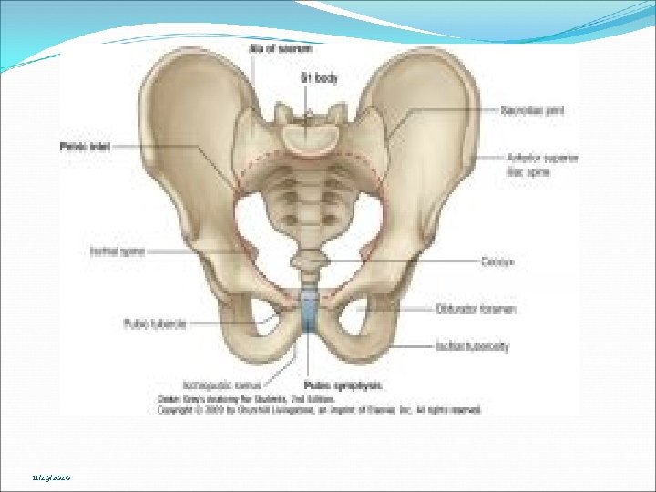
Other ligaments attached to bony pelvis include the sacrococcygeal ligaments, pubic symphysis ligaments, and endopelvic fascia ligament. • posterior boundary—sacrum and coccyx. Female pelvis with ligaments muscles and organs, 4 part. Each pelvic bone (hip bone) is made by the combination three bones namely, the ilium, pubis, and ischium. An array of issues can manifest themselves causing pain or dysfunction of the pelvis. The axial (horizontal) image of the female pelvis shows the ovaries, uterus, ligament, uterine tubes, vaginal cavity, and other internal organs. Additional ligaments may be found in the female pelvis. S4 cleveland clinic journal of medicine volume 72. Cookies allow us to analyze and store information such as the characteristics of your device as well as certain personal data (e.g., ip addresses, navigation, usage or geolocation data, unique identifiers). Surgical anatomy of the female pelvis. The broad ligament and the round ligaments of the uterus serve as a secondary support for the uterus within the pelvis. The broad ligament forms after the mullerian ducts join together during development. The cardinal ligaments lie at the base of the broad ligament and are described under the section on supportive tissues and cleavage planes.
In this section, learn more about the anatomy of the pelvis, and the. • posterior boundary—sacrum and coccyx. Each pelvic bone (hip bone) is made by the combination three bones namely, the ilium, pubis, and ischium. • anterolateral wall—hip bone and obturator internus muscles. These ligaments also play a crucial role in pelvic organ prolapse with anterior vaginal wall descent (5).
Pelvis Skeleton Model Female 6 Part 3b Scientific 1000288 H20 4 from www.galaxymed.de Therefore, the ligaments are essential components in the maintenance of a normal position of the uterus within the female pelvic cavity. • anterolateral wall—hip bone and obturator internus muscles. Many pelvic landmarks, ligaments, and muscular structures within the pelvis are important to know to differentiate normal reproductive organs from muscular and vascular structures. Ischial tuberosities, sacrotuberous and sacrospinous ligaments and, tip of the coccyx. An array of issues can manifest themselves causing pain or dysfunction of the pelvis. The pelvis is held together by three principal ligaments: Sports, trauma, pregnancy and a sedentary lifestyle are all contributors to this. Pelvic anatomy www.freelivedoctor.com slideshare uses cookies to improve functionality and performance, and to provide you with relevant advertising.
• muscles and ligaments form a pelvic floor.
The sagittal (longitudinal) image of the female pelvis shows anatomical structures. • anterolateral wall—hip bone and obturator internus muscles. The skin, tissues and organs in the pelvis are supplied by the vasculature of the pelvis, and innervated by many nerves of the pelvis, including the pudendal nerve. The right half of the model shows the following pelvic ligaments: See ligaments of the female pelvis below. In women, the ligaments of the joint soften during pregnancy, enabling the increase of pelvic diameter during childbirth. These ligaments also play a crucial role in pelvic organ prolapse with anterior vaginal wall descent (5). Interactive video showing normal female pelvic anatomy as seen by laparoscopy. The broad ligament can be further divided into three components that are linked to. An array of issues can manifest themselves causing pain or dysfunction of the pelvis. Additional ligaments may be found in the female pelvis. The pelvis is held together by three principal ligaments: The inlet to the pelvic canal is at the level of the sacral promontory and superior aspect of the pubic bones.
Pelvic anatomy sacrouterine ligament cardinal ligaments pelvic fascia sacrospinous ligament urethral support bladder support rectal support pelvic anatomy. S4 cleveland clinic journal of medicine volume 72.


0 Komentar